You may have heard about these little organelles — they are pretty in vogue now and are having their heyday. Interest in and research into mitochondria is constantly growing, and we are finding out just how important they are to the function of our cells and systems. What’s funny is that I learned all about them in undergrad and medical school, but I didn’t give them much thought once I became a doctor. However, I should be thinking about them a lot because the health of humans depends a great deal on them.
So, what are mitochondria?
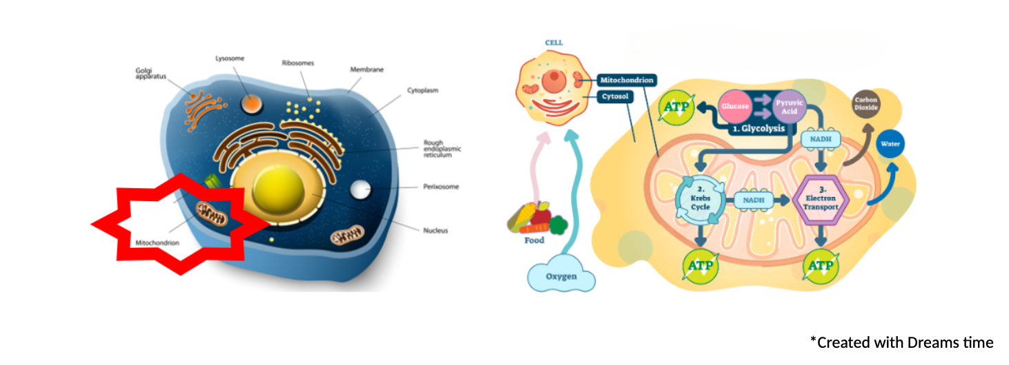
The mitochondria are tiny organelles found in nearly all our cells. But don’t let their small size fool you — the mitochondria are the cell’s powerhouses. One of their main functions is energy production. This is where fats, carbohydrates, and protein are converted into ATP (adenosine triphosphate), the fuel that powers the cell. No ATP = no energy = dead cell.
As mitochondria are essential for energy production, they are found abundantly in the organs that require the most energy — the brain, muscles, and heart.
In addition to ATP production, research has also discovered many other crucial roles the mitochondria play, including:
- Steroid hormone synthesis. The first step in creating steroid hormones (estrogens, testosterone, progesterone) occurs in the mitochondria!
- Heme synthesis. Heme is essential for creating hemoglobin and the cytochrome proteins that produce ATP in the electron transport chain.
- Cell death. Apoptosis, or programmed cell death, is a controlled process that removes damaged cells and maintains tissue integrity. The mitochondria act as a critical checkpoint in this process.
- Regulation of the immune response. The mitochondria produce energy for the immune system and regulate the signals within the immune cells that guide their activation and proliferation.
Phew! They are some hardworking organelles!
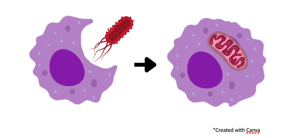
Nerd alert: The evolutionary history of mitochondria is fascinating! The most widely accepted theory is that billions of years ago, mitochondria were once free-living bacteria engulfed by a larger host. But, instead of being entirely digested by the host, it formed a symbiotic relationship; these engulfed bacteria produced energy in exchange for the host providing nutrients and protection.
Want to nerd out more? Check this out: https://youtu.be/39HTpUG1MwQ?feature=shared.
Ultimately, sustaining health means sustaining healthy mitochondria. Dysfunctional mitochondria lead to reduced ATP production and excessive free radical production, which can activate harmful cellular pathways. Unfortunately, it is pretty easy for the mitochondria to become dysfunctional.
- Aging. Like every other part of us, mitochondria age and get damaged.
- Reactive oxygen species (ROS) or free radicals. During regular operation, just like a car engine kicks off toxic exhaust, the mitochondria create ROS, which are highly reactive and can damage DNA, cell membranes, and proteins.

- Mutations in mitochondrial DNA. The mitochondria have their own DNA (remember they were once free-living bacteria?), but unlike the DNA in the nucleus, mitochondrial DNA is not as well protected and is much more prone to mutations and damage.
Fortunately, the mitochondria have systems in place to keep them functioning well.
- Antioxidant enzymes and antioxidants like glutathione. Antioxidant pathways and molecules quench the ROS and free radicals produced by the mitochondria. Deficiencies in these processes or molecules can allow the free radicals to cause damage.
- Fission, fusion, and mitophagy. Have you ever seen a lava lamp — where the little blobs join together to form a bigger blob or split off and become smaller blobs? The mitochondria do something similar but with intention. Damaged mitochondria may either fuse with other mitochondria to overcome any deficits or pinch off a damaged piece and tag it for destruction in mitophagy. Mitophagy is an integral part of quality control of the mitochondria and maintains mitochondrial efficiency. Mitophagy dysfunction has been found to occur naturally with aging and has been linked to chronic health conditions.
I tried to draw a picture of this process, but alas, my Canva skills were inadequate. Instead, check out this short clip from the University of Cambridge. https://youtu.be/tWP4XKTQi88?feature=shared
Despite the mechanisms to repair itself, given enough oxidative stress and insults, the mitochondria can sustain damage and become dysfunctional. You can imagine that if your mitochondria are not working well, this will have huge impacts on health, especially on the organs heavily reliant on energy production — remember, the brain, muscles, and heart. Here are just some of the diseases that mitochondrial dysfunction has been linked to:
- Brain Aging
- Parkinson’s disease
- Atherosclerosis
- Kidney disease
- Immune dysfunction
And, of course, we can’t talk about mitochondrial dysfunction without discussing COVID-19. More and more research is finding that the inflammation, apoptosis (cell death), and endothelial dysfunction seen in COVID-19 likely result from mitochondrial dysregulation that occurs with the infection. This sheds some light on the symptoms of long COVID — chronic fatigue, exercise intolerance, and brain fog… notice the affected organs — brain and muscles.
So, how can we support our mitochondria?
- Exercise. Regular exercise stimulates the creation of new mitochondria within cells.
- Nutrients! For our mitochondria enzymes to efficiently generate energy, besides getting enough carbs, fats, and proteins, they also need adequate cofactors. Some of these include:
- Thiamine (B1)
- Riboflavin (B2)
- Niacin (B3 — supplemental forms as NAD+ or NMN)
- Pantothenic acid (B5)
- Magnesium
- Iron
- Lipoic acid
- Coenzyme Q10
- Carnitine
We can obtain these nutrients from eating a diverse, healthy diet. Our body can also make its own Coq10 and carnitine. However, if we are deficient, we may need supplementation of these compounds.
- Poor diets — may be deficient in many of these compounds.
- Menstruating women — may be low in iron.
- Vegans or vegetarians — may be low in carnitine (the food source of carnitine is highest in animal products).
- Statin medication — statin medications block an enzyme that is also utilized in Coq10 production.
As with any supplements, please discuss them with your healthcare provider.
- Antioxidants. Remember all those free radicals produced by the mitochondria? Having sufficient antioxidants can help quench free radicals. Again, a diet comprised of various plants can be beneficial.
- Supplements. As highlighted earlier, you can supplement with nutrients if you are deficient, as well you can supplement with antioxidants. Another exciting supplement being investigated is urolithin A. Urolithin A is produced by the gut microflora from foods rich in ellagic acid and ellagitannins, such as those found in pomegranates, certain berries, and nuts, and it has been demonstrated to activate mitophagy and improve mitochondrial health. However, the ability to make urolithin A varies from person to person depending on their gut microbiome, and you would also have to eat a lot of pomegranates to get significant levels. Supplementation with this compound is still being researched, but it seems promising.
- Red light therapy! Check out my other blog to learn how red light stimulates the mitochondria.
- Cold exposure. The research on the effects of cold exposure on mitochondrial health is still growing and developing. However, so far, it seems that cold exposure may stimulate mitochondrial biosynthesis and improve the activity of mitochondria. It also increases the amount of brown fat; brown fat is a type of fat tissue that plays a role in heat generation and is associated with improved metabolic parameters. Currently, the optimal type or duration of cold exposure is still unclear. Cold exposure has risks, especially if you have underlying health conditions. Before plunging into this one, speak with your healthcare provider.
My take homes:
As always, focus on a healthy lifestyle –eat well, exercise, manage stress, and get enough sleep! However, if you have chronic health issues such as fatigue, cognitive decline, or heart or muscle issues, perhaps your mitochondria could use a boost. Ask your healthcare provider if they think any of these or other interventions may benefit (and not harm) you and your mitochondria!
Cheers to your mitochondria!
Further reading:
Mitophagy
Onishi M, Yamano K, Sato M, Matsuda N, Okamoto K. Molecular mechanisms and physiological functions of mitophagy. EMBO J. 2021 Feb 1;40(3):e104705. doi: 10.15252/embj.2020104705. Epub 2021 Jan 13. PMID: 33438778; PMCID: PMC7849173.
Brown Fat
Noriega L, Yang CY, Wang CH. Brown Fat and Nutrition: Implications for Nutritional Interventions. Nutrients. 2023 Sep 20;15(18):4072. doi: 10.3390/nu15184072. PMID: 37764855; PMCID: PMC10536824.
Mitochondrial Dysfunction and Diseases
Bondy SC. Mitochondrial Dysfunction as the Major Basis of Brain Aging. Biomolecules. 2024 Mar 26;14(4):402. doi: 10.3390/biom14040402. PMID: 38672420; PMCID: PMC11048299.
Xu M, Wang W, Cheng J, Qu H, Xu M, Wang L. Effects of mitochondrial dysfunction on cellular function: Role in atherosclerosis. Biomed Pharmacother. 2024 May;174:116587. doi: 10.1016/j.biopha.2024.116587. Epub 2024 Apr 17. PMID: 38636397. Zorov DB, Abramicheva PA, Andrianova NV, Babenko VA, Zorova LD, Zorov SD, Pevzner IB, Popkov VA, Semenovich DS, Yakupova EI, Silachev DN, Plotnikov EY, Sukhikh GT. Mitocentricity. Biochemistry (Mosc). 2024 Feb;89(2):223–240. doi: 10.1134/S0006297924020044. PMID: 38622092.
Yang K, Li T, Geng Y, Zou X, Peng F, Gao W. The role of mitophagy in the development of chronic kidney disease. PeerJ. 2024 Apr 25;12:e17260. doi: 10.7717/peerj.17260. PMID: 38680884; PMCID: PMC11056108.
Guo Y, Che R, Wang P, Zhang A. Mitochondrial dysfunction in the pathophysiology of renal diseases. Am J Physiol Renal Physiol. 2024 May 1;326(5):F768-F779. doi: 10.1152/ajprenal.00189.2023. Epub 2024 Mar 7. PMID: 38450435.
Borsche M, Pereira SL, Klein C, Grünewald A. Mitochondria and Parkinson’s Disease: Clinical, Molecular, and Translational Aspects. J Parkinsons Dis. 2021;11(1):45–60. doi: 10.3233/JPD-201981. PMID: 33074190; PMCID: PMC7990451.
Trinchese G, Cimmino F, Catapano A, Cavaliere G, Mollica MP. Mitochondria: the gatekeepers between metabolism and immunity. Front Immunol. 2024 Feb 23;15:1334006. doi: 10.3389/fimmu.2024.1334006. PMID: 38464536; PMCID: PMC10920337.
COVID-19 and mitochondria
Georgieva E, Ananiev J, Yovchev Y, Arabadzhiev G, Abrashev H, Abrasheva D, Atanasov V, Kostandieva R, Mitev M, Petkova-Parlapanska K, Karamalakova Y, Koleva-Korkelia I, Tsoneva V, Nikolova G. COVID-19 Complications: Oxidative Stress, Inflammation, and Mitochondrial and Endothelial Dysfunction. Int J Mol Sci. 2023 Oct 4;24(19):14876. doi: 10.3390/ijms241914876. PMID: 37834324; PMCID: PMC10573237.
Molnar T, Lehoczki A, Fekete M, Varnai R, Zavori L, Erdo-Bonyar S, Simon D, Berki T, Csecsei P, Ezer E. Mitochondrial dysfunction in long COVID: mechanisms, consequences, and potential therapeutic approaches. Geroscience. 2024 Apr 26. doi: 10.1007/s11357–024–01165–5. Epub ahead of print. PMID: 38668888.
Jenkins BC, Neikirk K, Katti P, Claypool SM, Kirabo A, McReynolds MR, Hinton A Jr. Mitochondria in disease: changes in shapes and dynamics. Trends Biochem Sci. 2024 Apr;49(4):346–360. doi: 10.1016/j.tibs.2024.01.011. Epub 2024 Feb 23. PMID: 38402097; PMCID: PMC10997448.
Urolithin A
Zhao H, Song G, Zhu H, Qian H, Pan X, Song X, Xie Y, Liu C. Pharmacological Effects of Urolithin A and Its Role in Muscle Health and Performance: Current Knowledge and Prospects. Nutrients. 2023 Oct 19;15(20):4441. doi: 10.3390/nu15204441. PMID: 37892516; PMCID: PMC10609777.
Cold Therapy:
Mahalingam S, Cheviron ZA, Storz JF, McClelland GB, Scott GR. Chronic cold exposure induces mitochondrial plasticity in deer mice native to high altitudes. J Physiol. 2020 Dec;598(23):5411–5426. doi: 10.1113/JP280298. Epub 2020 Sep 14. PMID: 32886797; PMCID: PMC8329962.
Mohan MS, Aswani SS, Aparna NS, Boban PT, Sudhakaran PR, Saja K. Effect of acute cold exposure on cardiac mitochondrial function: role of sirtuins. Mol Cell Biochem. 2023 Oct;478(10):2257–2270. doi: 10.1007/s11010–022–04656–1. Epub 2023 Feb 13. PMID: 36781815.
For more useful information on functional holistic health, you can visit our FREE video library here.
About the Author:
Dr. Eri Shimizu is a board certified in Internal Medicine Doctor and certified through the Institutes of Functional Medicine. She earned a Bachelor of Science in Environmental Bioengineering from the University of Hawaii at Manoa and graduated summa cum laude from Creighton University Medical School. She completed her Internal Medicine residency at UCLA and worked at a Los Angeles county hospital. In 2012, she returned to Hawaii and served as a Hospitalist at Maui Memorial Medical Center. Maui is now home with her husband, two children, and a fighting fish named Rainbow.
Schedule a FREE Functional Medicine Health Consult with Dr. Eri.